How the Patient Presented
The patient came in concerned about a tired, aged appearance in the periorbital area that she felt was disproportionate to the rest of her face. On examination, the clinical picture was defined by two distinct but interrelated structural problems.
The first was marked laxity of the lower eyelid structures — a progressive weakening of the supporting tissues that normally hold the eyelid in its correct, firm position. The second was herniation of periorbital fat: the fatty tissue that ordinarily cushions and protects the eyeball had migrated forward, pushing outward against the eyelid wall and forming visible, well-defined pouches beneath the eyes. Adding to this was a tear trough — a pronounced groove at the junction between the eyelid and the cheek — that deepened the contrast in volume and intensified the shadow under the eye.
The Treatment Decision
The complexity of this case required a tailored surgical approach capable of addressing all three components simultaneously — excess volume, volume deficit, and structural laxity — without compromising the naturalness of the final result.
Simple fat excision would have partially resolved the pouches but would have deepened the tear trough further, creating an excavated, unnatural appearance. A redistribution-only approach, without excision, would not have eliminated enough excess volume. The decision was to combine both techniques and complement them with reinforcement of the eyelid’s supporting structures.
Surgical Technique
The procedure of lower blepharoplasty consisted of:
- Selective excision of herniated fatty tissue. The precise amount of fat responsible for the prominence of the pouches was removed, while preserving the volume needed for the next step. Accuracy here is critical — over-excision produces a hollowed appearance that is difficult to correct afterward.
- Fat redistribution into the tear trough. A portion of the excised fatty tissue was repositioned and secured into the tear trough zone, filling the depression at the eyelid–cheek junction. This is the step that ensures a smooth, shadow-free transition with no abrupt change in volume — the key element of a truly natural result.
- Reinforcement of the lower eyelid supporting structures. The residual eyelid laxity was corrected by consolidating the supporting tissues, restoring correct position and tone to the lower eyelid. Without this step, the aesthetic result would have been compromised in the medium term as laxity re-emerged.
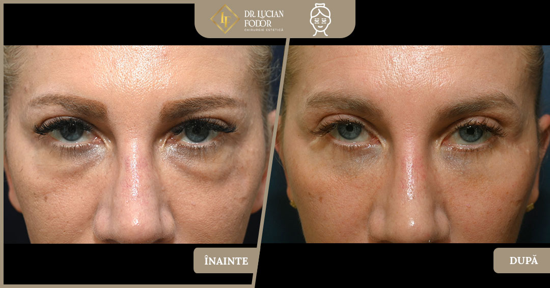
Result
The periorbital area now presents a uniform contour — no pouches, no pronounced tear trough, and no sagging eyelid appearance. The gaze is more open and brighter, and the transition between eyelid and cheek is smooth and natural. The outcome is in harmony with the rest of the face — with nothing to announce a surgical procedure.